A newly-discovered niche for Polyarthra vulgaris?

So, seeing as I’m pre-writing this post at the moment, these finds are about a week and a half old, but no less awesome.

As of writing this, my puddle water sample from Renfree Field in Sacramento has been aging for about 4 or 5 days. Over the course of this time, the water’s temperature, lighting, pH, turbulence and bacterial population have changed enough in a well-lit corner of my bedroom-turned-microbiology lab (relative to a puddle in an outdoors parking lot) such that the normally-dominant species in nature no longer have the competitive advantage they did when I first collected my sample. What is very interesting is that this means some species which normally exist in only tiny populations in the wild then have a chance to seize the newly-vacated water column and grow to high enough densities that I actually find them. In the case of rotifers, the primary species Epiphanes senta and perhaps Asplanchna silvestrii die out to yield Brachionus of some kind. And now, as I’ve continued my observations, I’ve found another late bloomer: Polyarthra vulgaris. 

Polyarthra_vulgaris
Polyarthra vulgaris from puddle water, imaged at 100x.

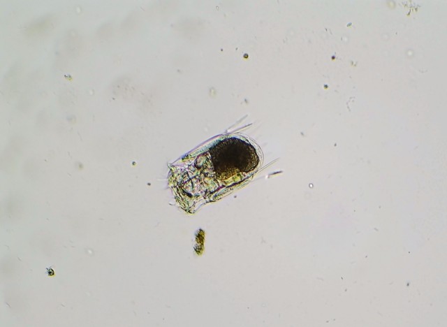
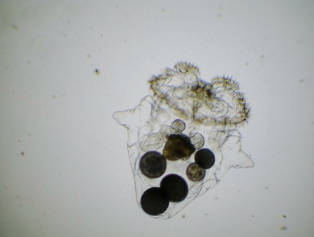
P. vulgaris is a planktonic, roughly square-shaped, soft-bodied, and relatively small rotifer in comparison to the ones I’ve previously featured on my blog – probably no more than 100 microns on average in any dimension. However, it makes up for its lack of size with a very feisty character, if one could anthropomorphize a rotifer in such a manner. This is mostly due to Polyarthra‘s unique set of swimming fins or paddles – pictured as the “spines” running down the side of the animal’s body – which it can flick very rapidly to “jump” through the water column. P. vulgaris thus has a two-speed swimming mechanism: a slow, steady and controllable cruising speed, with motion generated by the regular rotation of the cilia which the rotifer uses to eat; and the quick, random and near-instantaneous jump through the water, a result of the lighting-fast twitch of the swimming fins. Many zooplankton species exhibit these two different types of swimming, especially the species on the lower end of the food chain. The lightning jump likely developed due to a need to escape from predators (say, a hungry Asplanchna), but as it is random and costs lots of energy, the organism still uses its regular feeding motion to move around slowly and more purposefully when it is not in immediate danger. (Just another aside on the lightning jump: in a normal drop of water, these rotifers legitimately appear to teleport. That’s how fast they are.)

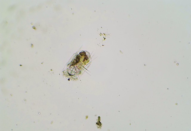
And here’s a slideshow of three frames I captured of the rotifer demonstrating its flicking motion:

This slideshow requires JavaScript.

And a video:

While the genus Polyarthra, to my knowledge, has been studied phylogenetically with DNA evidence and no doubt contains hidden species like the Epiphanes senta case, it’s still possible to identify general species “groups” based off of the visible structure of the swimming fins. Using a simple dichotomous key for zooplankton I found online, I am tentatively calling my individuals P. vulgaris due to their generally short, sleek fins. This species was first described in 1943, in a German book titled Die Plankton Rotatorien des Motalastrom: zur Taxonomic and Okologie der Plankton Rotatorien; it is a close relative of the related and longer-finned species P. dolichoptera, with which it often grows in the same body of water. The majority of research I’ve found on Polyarthra concerns its population dynamics, specifically with regards to predator-prey relationships and interspecific competition. In looking at the head-to-head comparison of the two aforementioned species of  PolyarthraP. vulgaris seems to have an advantage in warmer and more oxygen-rich waters (corresponding with the highest populations in summer and autumn), whereas P. dolichoptera prefers water up to 15 degrees Celsius cooler and can tolerate lower levels of dissolved oxygen (which explains why it often reaches highest population densities in the spring). However, both species can proliferate well in water below 10 degrees Celsius and will survive below 5.

So here’s where things get interesting.

Polyarthra_vulgaris_2
Another individual of Polyarthra vulgaris, imaged at 100x.

As far as I know, P. vulgaris has been observed and collected from lakes and ponds in the northern parts of the continental United States and Europe, with one aberrant habitat being a canal in Washington. It can inhabit both oligotrophic (nutrient-poor) and eutrophic (nutrient-rich) waters, suggesting that it is relatively adaptable given its preferred temperature conditions. However, I have not come across in my reading any records of Polyarthra from ephemeral waters such as vernal pools or in this case a puddle. Knowing that temperature is often a major factor in their competitiveness and the rotifers’ recorded locales, I suspect that Polyarthra species must have a dormant phase during which they diapause or hibernate when the water warms up during the summer (especially in the case of P. dolichoptera, which prefers water temperatures of only 3-5 degrees Celsius!), and this has been reflected in the seasonal changes in these rotifers’ densities. Thus I wouldn’t be surprised if this population has that ability to diapause and regenerate, but it is pretty awesome to discover a potentially never before recorded habitat for them – in which they can not only survive unfavorable changes in temperature but also desiccation.

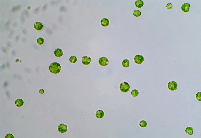
So that’s a wrap on Polyarthra vulgaris, with a brief overview of its unique morphology, research interest and what I found interesting about it. I’ll probably have a few more posts on puddle-water microbes (and of course lots of stuff on Chlamydomonas) before I switch topics for a little while. Stay tuned.

Works cited:

Allen, A. A. (1968). Morphology of the Planktonic Rotifer Polyarthra vulgaris. Transactions of the American Microscopical Society, 87(1), 60-69. doi:10.2307/3224338

Haney, J.F. et al. “An-Image-based Key to the Zooplankton of North America” version 5.0 released 2013. University of New Hampshire Center for Freshwater Biology <cfb.unh.edu> 8 Dec 2017

Laxhuber, R. (1987). Abundance and distribution of pelagic rotifers in a cold, deep oligotrophic alpine lake (Königssee). Hydrobiologia, 147(1), 189-196.

Saunders-Davies, A. (1989). Horizontal distribution of the plankton rotifers Keratella cochlearis (Bory de St Vincent) and Polyarthra vulgaris (Carlin) in a small eutrophic lake. Hydrobiologia, 186(1), 153-156.

Stenson, J. (1983). Changes in the relative abundance of Polyarthra vulgaris and P. dolichoptera , following the elimination of fish. Hydrobiologia, 104(1), 269-273.


The Green Yeast, Part 2 – the basic anatomy of Chlamydomonas reinhardtii

In my previous post, I introduced the alga Chlamydomonas reinhardtii as a small, single-celled species that lived in puddle water. Which probably narrows down the possibilities to maybe several hundred algae and several thousand protozoa, so in this post, I figured I’d flesh out that description a little more.

In the future, I’ll cover the phylogeny and taxonomy of C. reinhardtii in more depth, but for now it’s relevant to understand that this alga belongs to the order Volvocales. Why? Well, it is generally well understood that chlamy is an ancestor or a progenitor form of the colonial Volvocales species I’ve discussed in previous posts, and it so happens that a lot of the cellular characteristics of PleodorinaEudorina, Pandorina and Volvox are also shared with Chlamydomonas. Let’s dissect one image of these cells, with arrows pointing to different structures of the alga in each repeated frame:

Chlamydomonas_anatomy
An assortment of Chlamydomonas reinhardtii, imaged at 400x.

The majority of normal vegetative cells of Chlamydomonas look like these: small, round, and solitary. They range in size from around 5 to 40 microns in diameter based off of my casual measurements with an eyepiece reticle of wild-type cells, although I would guess that most of them are closer to 20 than either of the other extremes.

Chlamydomonas_exopolysaccharide
Chlamydomonas reinhardtii, with arrows pointing to exopolysaccharide secretions. Imaged at 400x.

The border of each cell which defines it from the extracellular milieu is a cell wall – a generally-tough outer layer which helps protect the cell beyond the lipid membrane which is present in all living organisms. While most animal cells do not have such a cell wall, many fungi, plants and algae do. In most plants, the cell wall is comprised almost entirely of cellulose, an example of compounds called polysaccharides (literally “many sugars”) which are composed of sugar molecules closely linked together. We encounter cellulose as both dietary fiber and in most paper or plant fiber products we use every day. The cell wall of Chlamydomonas, on the other hand, has a high proportion of glycoproteins and glycopeptides, which are special types of proteins with sugars embedded in their structure. Another component of the Chlamydomonas cell wall is polysaccharide, but rather than forming stringy fibers like cellulose does, these compounds are very gel-like. In some strains of Chlamydomonas, a faint, clear ring of this gel is also produced outside of the cell wall and can be seen encircling the pigmented cell – here, a few cells with this form are notated with arrows, although it is fairly difficult to see. This material is known as extracellular polysaccharide, or exopolysaccharide for short.

Chlamydomonas_chloroplast
Chlamydomonas reinhardtii, with arrows pointing to cup-shaped chloroplast. Imaged at 400x.

Within the cell now…all healthy Chlamydomonas individuals have this vibrant light green color. However, it is clearly not evenly distributed; the most saturated color can be found circling the bottom and sides of the cell wall in a thin layer. This “u”-shaped or cup-shaped structure is the cell’s chloroplast, and it is the major light-harvesting organelle in Chlamydomonas. Its green color comes from different forms of the compound chlorophyll, which harvest light energy and help the cell synthesize its own sugars to turn into energy for swimming, growth and reproduction. Although it does not take up the majority of the cell’s volume, the chloroplast covers much of the inner surface area to maximize the light that each cell can harvest.

Chlamydomonas_pyrenoid
Chlamydomonas reinhardtii, with arrows pointing to pyrenoid. Imaged at 400x.

You may have noticed that in many of these cells, there appears to be a bulge in the chloroplast, and in the indicated cells this bulge is shown to be its own distinct outlined structure. This is the pyrenoid, and as is evidenced by its proximity to the chloroplast, it plays a very important role in photosynthesis. As some of the research which I am very interested in concerns the pyrenoid, I will save the detailed description of its function for a future post. For now, it’s probably easiest to remember that it is an integral component of the photosynthesis process in Chlamydomonas, particularly the steps in which carbon dioxide is turned into cellular sugars.

Chlamydomonas_eyespot
Chlamydomonas reinhardtii, with arrows pointing to eyespot; note the faint red color. Imaged at 400x.

The lighter-colored section of the cells is the cytoplasm, containing the gelatinous fluid which gives the cell its volume (it should be clear, but the chloroplast is three-dimensional and covers part of the view into the cell). The eyespot of Chlamydomonas is usually located here, and it is the secondary light-harvesting organelle in this alga. Unlike the chloroplast, however, the eyespot is not used for generating cellular energy; it instead can sense changes in the concentration of light, communicating with the rest of the cell to direct movement and maximization of photosynthesis. Chlamydomonas thus demonstrates phototaxis, or movement in response to light, specifically positive phototaxis or movement toward light (for the benefit of increased photosynthesis and cellular energy). Although it’s hard to see, these eyespots have a reddish-orange color to them – why is this? Well, have you ever heard that Vitamin A is good for the eyes, and carrots have lots of Vitamin A? While this is an oversimplification, the short story is that Chlamydomonas eyes need Vitamin A to function properly too, and this reddish color comes from the Vitamin A in its active form, concentrated in a single organelle.

Chlamydomonas_contractile_vacuole
Chlamydomonas reinhardtii, with arrows pointing to contractile vacuoles. Imaged at 400x.

While the cells are spherical and it’s difficult to assign them a “head” or “tail”, the “head” of Chlamydomonas is generally the side on which this feature appears. These tiny, clear spherical regions in the cell are contractile vacuoles, and are very common among protozoa as well as algae. Their role is very simply to pump water out of the cell. But wouldn’t this end up drying out the cell very quickly? No, thanks to the process of osmosis. Osmosis is a special type of diffusion, where water crosses a permeable membrane to try and reach an equilibrium – where the concentration of solutions on either side of the membrane are equal. For example, if a cell has lots of dissolved sugars, proteins and salts in its cytoplasm, it will have a higher concentration of solutes in it than the surrounding water has. To try to reach an equilibrium, water will preferentially move across the cell membrane into the cell, diluting its contents until the concentration of solutes is the same inside and out. In their natural environment, most single-celled aquatic organisms (Chlamydomonas included) like to maintain this positive water balance, where water flows into the cell constantly, because it is easier to pump water out than to try to suck it back in. To prevent the cell from exploding with water pressure, contractile vacuoles are used to pump extra water out; as the water flows back in, the contractile vacuoles inflate and deflate at regular intervals. This costs the cell a constant expenditure of energy, but it is essential for their survival. Most cells of Chlamydomonas have two contractile vacuoles, and they are almost always located together at this “head” of the cell.

Chlamydomonas_flagella
Chlamydomonas reinhardtii, with arrows pointing to exopolysaccharide secretions. Imaged at 400x.

Another organelle which helps define the head of the cell are these faint protrusions which have their base near the contractile vacuoles and extend outwards like little hairs. These are flagella, and they are the primary mode of locomotion for Chlamydomonas. Like the whips for which they are named (in Latin), flagella sweep back and forth rapidly in the water, creating a current which the cell uses to swim. Mature and healthy cells of Chlamydomonas have two of these each, but they often drift in and out of focus as the cell moves them around and are thus hard to see in every individual; additionally, they may fall off of older cells or cells that have been subjected to lots of physical handling (like being squeezed by a coverslip on a microscope slide, for instance).

So that’s a basic overview of the visible structures of Chlamydomonas using light microscopy. Although there are of course more – a nucleus, mitochondrion, Golgi body – the six that I covered here (seven if you count the cytoplasm) are very heavily studied and I will discuss these in depth in the future, so I thought it would be a good review for me to go over their functions and cellular locations – and hopefully interesting for you as well. My next few posts will not be Chlamydomonas-related – I have some other puddle water organisms to share – but I will be sure to continue this series, especially as I explore possible areas of research for this little alga. Stay tuned.


The Green Yeast, Part 1 – Chlamydomonas reinhardtii glory shots

So now, I’ve finally arrived at this single-celled alga, Chlamydomonas reinhardtii, affectionately called “chlamy”, which I’ve observed in puddle water alongside the more spectacular colonial species.

This being probably the single most heavily-studied alga in history, I felt it deserved itself a good series of blog posts to cover all of the different aspects of its biology, history, applications and my personal connections to it.

But before then, here are some of the shots I’ve managed to capture of C. reinhardtii in situ, or in its natural habitat:

Chlamydomonas_montage
Chlamydomonas reinhardtii from puddle water, imaged at 100x.

Chlamydomonas reinhardtii is quite numerous in many of the puddles that Eudorina and Pandorina also inhabit, and like those two algae it tends to swim towards and cluster in the most strongly-lit area of a sample. This picture was taken at one end of a drop of water after about 2 minutes of letting it sit on the microscope slide, after which all of the chlamy had swarmed to that end, stopped swimming so erratically, and were easier to photograph.

Chlamydomonas_montage_2
Chlamydomonas reinhardtii, imaged at 400x.

As is evident at the closer magnifications, the cells of chlamy aren’t just uniform blobs of green (some species of algae unfortunately do just look like that, and they’re an absolute pain to try and identify by eye alone). In my next post, I plan to cover the basic anatomy of a cell of C. reinhardtii so that these features become more obvious and meaningful as I continue my discussion of the alga. Then, I’ll do an in-depth research post to dig up some of the history of C. reinhardtii. After that, I’ll cover some of the biggest areas of biology in which chlamy is currently being used, hopefully one post per topic. And finally, I’ll legitimately discuss my lab work for the first time on this blog as I show my own cultures of this alga and explain in brief my plans for them, both currently and as I move into college (the latter is of course subject to change). In between these posts, I’ll sandwich in some other interesting finds from my puddle water samples, including the colonial alga Gonium and hopefully the giant ciliate Bursaria truncatella. Lots of good stuff planned for the next couple of weeks. Stay tuned.

SLIME VIDEO except alive and it has a shell – testate amoebae from puddles

So as I’ve been continuing to survey puddle water from now multiple locations (having collected some more from a new, muddier puddle in a parking lot), I’ve discovered more and more awesome organisms. These include Eudorina in huge numbers (thousands in a single drop of water), as well as other types of colonial algae and some more rotifers.

But by far my favorite (non-algal) discovery in puddle water has been testate amoebae.

Arcella
A small, buoyant individual of what appears to be Arcella sp. collected from puddle water, imaged at 100x.

When one says “amoeba”, the first thing that comes to mind is probably this clear, shape-shifting blob that spawns in the most disgusting of pond scum, morphing itself into the semblance of a giant mouth that eats anything that gets in its way. And by and large, that image is mostly correct. The term “amoeba” can refer to most any single-celled organism that can willingly and readily slosh around its cytoplasm (its internal jelly-like volume) and change shape. In doing so, it can not only eat (by surrounding its prey with its own body and engulfing it) but also move by grabbing onto objects and pulling itself towards them. This action is typically performed by extending jelly-filled projections out of the cell called pseudopods, which derives its etymology from the Greek for “false feet”.

While amoeboid organisms or organisms with amoeboid cells or life stages (including humans – the white blood cell called the macrophage travels through the blood and eats up invading bacteria and viruses!) are common and evolutionarily very successful, some problems with adopting a “blob” form quickly arise. The most obvious, it would seem, is the fragility of the organism – just stab it and it will pop, right? At the cellular level, it’s not that simple (I would presume – I know very little about amoebae), but clearly, as amoebae are usually slow and they must be flexible to change shape, they would appear to make easy prey.

One solution that amoeboid organisms have come up with is to create a shell, also known as a test – these particular amoebae are thus called testate amoebae. Like a hermit crab’s stolen shell, the test provides the amoeba with an element of protection, and can also be filled with air or water to help the amoeba sink or float, depending on its food source or need for light. Also like how a hermit crab can still poke out of its shell a little to collect food and move around, testate amoebae extend pseudopods from an opening in their test, which they use for hunting and locomotion. Testate amoebae are so successful that they have appeared in several unrelated lineages of microorganisms – the Arcellinida, a lineage of microorganisms that have practically exclusively testate amoeboid forms; the Rhizaria, which include saltwater plankton species as well as shelled amoebas; and the Stramenophila, many members of which (such as diatoms) are species of algae, and others of which are testate amoebae.

Testate_amoeba
An unidentified benthic testate amoeba from puddle water, imaged at 100x.

Although it’s relatively easy to find testate amoebae in most samples from permanent bodies of water, either tangled up in masses of algae or resting in layers of sediment (peat bogs are especially reputable places to look for these), I was quite surprised to see them in puddle water. However, some amoebae such as the infamous brain-eater Naegleria fowleri have multiple life cycle stages, including a cyst which is resistant to environmental hardship; thus, it’s not a stretch to imagine that other species can survive drying out completely.

I found two species in the puddle water: the first one pictured in this post is probably a representative of the common genus Arcella, and the second is unidentified. The small species of Arcella I typically see are normally an absolute pain to photograph because it is a buoyant species, meaning it likes to store gas bubbles inside its shell and float on the water’s surface. This behavior has been noted as early as in the 1910s, and while it may help the amoeba to collect its food, it means that it is prone to being blown around by tiny air currents and is thus impossible to take time-lapse video of. In addition, they usually like to hold their blobs of cytoplasm to themselves closely, without extending the pseudopods one expects from an amoeba. However, I found a single individual which behaved exactly the opposite – sinking to the bottom of the microscope slide and crawling around with classic amoeboid behavior – and I had to capture it on video. So here is a time-lapse of its motion at 10x real speed and 100x magnification:

On the other hand, the second unidentified amoeba species seems to always behave very well, sinking straight to the bottom of the water drop and extending several pseudopods. I presume that it prefers to search the sediments for edible particles, and its shell appears to be weighted down by mineralized material to give it additional weight and prevent its disturbance by water currents or wind. Sadly, whatever it is, I don’t think I will ever be able to find out. Several studies have found that they are practically impossible to culture, and I don’t have the right tools to sequence and analyze its DNA. But here is a video of it all the same, also filmed at 10x real speed and 100x magnification over the course of 3 minutes:

So that’s the basics on testate amoebae from puddle water. In future posts, I hope to find more of these from different bodies of water and share them here too. Stay tuned.

Works cited:

Bles, E. J. (1929). Arcella. A Study in Cell Physiology. Journal of Cell Science, S2-72, 527-642.

Gomaa, F., Kosakyan, A., Heger, T. J., Corsaro, D., Mitchell, E. A., & Lara, E. (2014). One Alga to Rule them All: Unrelated Mixotrophic Testate Amoebae (Amoebozoa, Rhizaria and Stramenopiles) Share the Same Symbiont (Trebouxiophyceae). Protist, 165(2), 161-176. doi:10.1016/j.protis.2014.01.002

Vegetative asexual reproduction of colonial Volvocales – an overview

In my previous posts, where I introduced the dominant colonial algal species I’ve seen in puddle water, I mainly discussed the appearance of their adult forms, only briefly mentioning that their reproductive stages are occasionally used to aid in their identification. However, as I’ve managed to capture a bunch of pictures of reproducing algae, I figured it would be interesting to post them here and give a general overview of their asexual reproduction.

The algae in the family Volvocaceae (well-known genera include EudorinaPandorinaPleodorina and Volvox) reproduce asexually by the production of identical colony units within their mother colonies. These “mini-colonies” develop within the mother colony until the mother ages and splits open; the minis are then released, grow larger, and develop smaller colonies inside them too, continuing the cycle. In a way, this could be considered a type of live birth, which is fairly common in algae; zoospore is the preferred name for motile, reproductive units produced by a parent (usually as a product of a parent colony’s cell) and released. Autospores are similar, with a common distinction being that they are a little smaller and usually non-motile. As reproduction is asexual, the daughter colonies are identical in DNA composition (with the exception of mutations), so when an entire population is formed consisting of individuals created through asexual reproduction, it is known as a clonal colony. 

Mature_colonial_Volvocales
Several mature colonies of Eudorina unicocca and Pandorina morum, the two most numerous colonial Volvocales in the puddle water I’ve sampled. Imaged at 100x.

So the lifecycle begins here, with mature colonies of the algae. Typically, one first notices algae in a puddle by their color, signaling that there is already a large population of actively-growing individuals, so scenes like these are pretty typical in initial observations of a sample.

Dividing_Eudorina
Eudorina sp. in early stages of asexual reproduction, imaged at 100x. Notice how not all cells are dividing synchronously.

Slightly older colonies typically become obvious within a day or two of letting a collected water sample sit in its container. In the individual of Eudorina pictured above, the vegetative cells have started to divide. Many of them have made one division, yielding two half cells; a couple (out of focus) have divided twice, yielding four cells. What is interesting is that these divisions do not yield new, identical cells. Rather, in their first couple of divisions, the resulting cells put together keep the shape of the original spherical cell, appearing as a semicircle and then a slice of pie, respectively.

Eudorina_daughter_colonies_1
A 16-celled colony of Eudorina, with 16-celled daughter colonies forming inside. Imaged at 100x.

As the mother colony grows older, the cells inside continue to divide, forming what will later become daughter colonies. By the time they have 16 cells, these colonies will have lost the spherical outline of the original cell, and instead flattened out into this square shape.

Eudorina_daughter_colonies_2
Another mature colony of Eudorina with daughter colonies developing inside. Note the square colony shape and the wide spacing of the daughter colonies inside the outline of the mother. Imaged at 100x.

In some colonies like this one, the actual boundaries of the colony expands as the daughter colonies develop inside it.

Eudorina_daughter_colonies_3
A species of Eudorina (probably not E. unicocca) with fully-formed daughter colonies. Imaged at 100x.

And mother colonies like this one pictured above are exceedingly rare, from what I’ve observed in natural samples. As the mother ages, the daughters can technically fully develop into small units identical to a mature adult, sort of like nymphs in some insects like grasshoppers. However, in most cases, the mother colony breaks open before it gets this old, and releases the daughter colonies while they are still in their “square” stage of development. By pipetting a couple of Eudorina colonies into a small tube and letting it sit undisturbed for a few days, I managed to induce these really unique mother colonies with advanced daughter colony development. As the mother colonies could just stay suspended in the water column without being pushed around by wind or water currents, they never split open and released the daughters.

Eudorina_releasing_daughters
A colony of a different species of Eudorina, in the process of splitting open. A few daughter colonies can be seen drifting away, as well as some cells which did not divide to produce daughter colonies. Imaged at 100x.

Finally, even the most delicately-preserved mother colonies age to a point where the gelatinous sheath of their colony breaks open. Sometimes, the daughter colonies even assist in this process, as they develop flagella and start jiggling inside the mother colony, perhaps adding some stress to its gelatinous matrix. When this split occurs, the daughter colonies are free to go, and swim off out of the mother colony to mature themselves, perpetuating the cycle.

A few final notes on asexual reproduction in Volvocales:

1) This article covered the process using pictures from different species of Eudorina, and it looks fairly similar in the closely-related Pandorina too; in fact, some of the above snapshots may have been Pandorina individuals, but I have no way of knowing for sure. In Pleodorina, it works slightly differently; as Pleodorina has two cell sizes, only the large cells divide to form daughter colonies. (See my post on Pleodorina californica for a few pictures of what this looks like.) Volvox reproduce with the same mother-daughter division system, but an even smaller proportion of Volvox cells form daughter colonies. Out of the thousands of cells which comprise a single colony, only a maximum of 5-10 on average will form daughter colonies, and these are specialized reproductive cells not used for locomotion or colonial structural integrity. Most mature Volvox usually already have daughter colonies formed, and it is very rare to find a colony of Volvox without any daughter colonies. In fact, the very old Volvox sometimes have “granddaughter” colonies, where their daughters are already developing daughter colonies inside them!

Volvox_with_daughters
Two mature individuals of different species of Volvox. The one on the left, imaged at 40x, has 8 daughter colonies; the one on the right is imaged at 100x and has 2 daughters.

2) When the algae sense vicissitudes in their environmental conditions, such as the drying up of a puddle, they can form a different reproductive body via asexual reproduction called an aplanospore. In this process, each vegetative cell does not start dividing like pictured above, but rather stays singular, turns dark brown to orange in color, and forms a thick cell wall. The mother colony dries out or breaks apart, and the cells fall out. These single-celled aplanospores can survive being dried out, and when environmental conditions become favorable again, they “hatch”, dividing and growing out into a full colony again. However, I’ve only seen this process work in nature (the puddles); when my algae grow old, they make aplanospores but they never hatch out again. I’ve seen a few Japanese papers describing new species of colonial Volvocales, and they basically say the same thing, that they haven’t been able to germinate their aplanospores in captivity. However, it would be really cool if I could – then I could keep like a little “seed bank” of Eudorina and grow it out any time I needed to for an experiment.

So that’s a general description of asexual reproduction amongst colonial members of the order Volvocales. Stay tuned.

The “Green Blackberry” – a profile of Pandorina morum (and very briefly, other Eudorina)

In my previous writings, I covered in depth the scientific history of, as well as my personal history seeing two colonial species of algae in the order Volvocales: Pleodorina californica and Eudorina cf. unicocca, both of which I’ve found in puddle water. In this post, I’ll quickly cover another colonial species in the genus Eudorina that I’ve seen, as well as a species in the related genus Pandorina.

My last post covered some of the defining features of Eudorina unicocca, including the round shape of its cells and the “5-row” arrangement of the cells in its colonies. However, not all Eudorina look like that. Here’s a couple of small individuals which have a different cellular morphology:

Eudorina_sp
Unidentified Eudorina sp., imaged at 100x.

Although the picture quality isn’t great, the bottom colony clearly doesn’t resemble the average Eudorina unicocca colony. Its cells aren’t round but rather teardrop-shaped, tapering to a point near the border of the colony. In addition, there doesn’t appear to be a particular arrangement of the cells within the colony, although to be fair, the colony is probably young and likely will take on a different appearance when it matures (however, I suspect that it will not get much bigger than this size). Each cell has inside it a single large pyrenoid – so does E. unicocca, but that alone is not enough to identify the species, and I believe that is some other species of Eudorina. I think that there might be several others besides the E. cf. unicocca I’ve seen, but I really won’t be able to confirm that unless I can start cultures of all of them, do more detailed microscopy and run DNA tests.

And now, the main focus of this post is another colonial alga, also of the order Volvocales and family Volvocaceae, but of a different genus, Pandorina. It’s quite closely related to Eudorina, and on first glance (at 40x, when surveying the water sample) it may be pretty hard to distinguish. However, when examined more closely, several features of this algae should appear very different from Eudorina unicocca:

Pandorina_morum
A mature individual of Pandorina cf. morum, photographed at 400x.

The first difference is the cell morphology. The cells of Pandorina are bigger. Much bigger. In addition, they’re not as round as the cells of most Eudorina, instead taking on a sort of geometric shape as they are compacted together. The cell surfaces, while not necessarily connected to their neighbors, are very close and in some cases do contact bordering cells. While it’s not as evident in this picture, some Pandorina individuals really have the appearance of “orange slices” in terms of just how tightly packed their cells are, which is very different from many of its relatives. However, in terms of cellular components, it’s pretty similar to the average cell of any alga in the order Volvocales (I’ll explain why this is in a future post): cup-shaped chloroplast (although it’s hard to see here, as the cells overlap each other in focus), single large pyrenoid (several are visible in this picture), an eyespot, and two flagella. The colony structure is fairly similar to that of Eudorina: round, generally oval in shape, consisting of a gelatinous matrix, with one smooth front end, one flattened back end, and a cell count of either 8, 16, or 32; five rows of cells, as described in E. unicocca, are somewhat discernible here. I haven’t run any DNA tests on this species, and I really don’t know how to identify it microscopically (or if that would even be a reliable method of identification), so for now I will just assign my observed individual the type species designation, Pandorina cf. morum.

The history of Pandoria morum is only noteworthy thanks to an interesting discussion of its etymology on its page on AlgaeBase, a website dedicated to cataloging species of algae and their taxonomic history online. P. morum got its current genus designation from a French publication in 1827 titled Encyclopédie méthodique ou par ordre de matières. Histoire naturelle des zoophytes, ou animaux rayonnés, faisant suite à l’histoire naturelle des vers de Bruguière, in which the algae was named Pandorina mora. However, this was not the first time the alga had been described. Forty years earlier, Otto Friedrich Müller (whom I’ve already mentioned in my post on Epiphanes senta) had seen this species, naming it Volvox morum in his work Animalcula infusoria fluviatilia et marina, published in 1786. Of course, he did not know that there existed other genera of colonial algae besides Volvox, so that revision was only made with the continued discovery of other species and revision of taxonomy. What was noteworthy about the alga’s description – or so holds M.D. Guiry of AlgaeBase – was not the change of genus assignment but the choice of species epithet, morum or mora. Upon consulting a Latin dictionary, Guiry found that the term “morum” could refer to either a mulberry or blackberry fruit; “mora” the whole plant; or either of the two an adjective meaning “silly or foolish” (from which the term moron originated, I assume). The logical conclusion, of course, is that morum, describing the algae like the fruit, was Müller’s true intent when naming the algae, so since then, the species has had its name restored to Pandorina morum, with the correct genus and the original epithet. (Although wouldn’t it be fun to name your algae the idiot algae… “Hey, have you published recently?” “Yeah, just described a new species.” “Oh cool, what’s it called?” “Oh, you know….Pandering moron.” Really gives science its necessary levity- I mean gravity.)

Pandorina_morum_2
Two young individuals of Pandorina cf. morum, imaged at 100x.

Last year, when I sampled the water from the same puddle, I found none of this alga, only the Eudorina cf. unicocca I described in my previous post. So this year, it was a nice surprise to see that it had not only appeared but veritably taken over the Eudorina and become the dominant species in my first few collected samples. However, I found that they seemed to be rather more sensitive and less hardy than the Eudorina, in the sense that after a couple of days, most of the Pandorina died in my samples while the Eudorina persisted for a little longer. In addition, as I collected more water in late November, I could no longer find very many or mature individuals of Pandorina, and Eudorina became the dominant colonial algae in the puddles once again. Obviously some factor must contribute to one’s competitive advantage over the other; what exactly that is, however, I don’t know. They seem to occupy similar niches in nature – same size, colonial structure, they both swim around in temporary bodies of water. I’d be interested in trying to find out what conditions each genus prefers by starting off two pure cultures of Eudorina and Pandorina, mixing them together in equal numbers in different environments, and checking ever so often to see what proportions of each alga can be found in the water. But that’s a lot of work and time, and I don’t know if I can even grow Pandorina, so I probably will save that experiment for some other time.

In my next post, I’ll discuss the life cycle of colonial algae, with more pictures; then, I’m thinking I can fit in one short piece on testate amoebae before I begin a long discussion on Chlamydomonas reinhardtii. Stay tuned.

Works cited:

M.D. Guiry in Guiry, M.D. & Guiry, G.M. 2017. AlgaeBase. World-wide electronic publication, National University of Ireland, Galway. http://www.algaebase.org; searched on 08 December 2017.


Another Californian alga, Eudorina cf. unicocca (including brief notes on G.M. Smith)

Eudorina cf. unicocca, as I have managed to identify it, is one of the dominant colonial algae that I’ve found in my puddle water samples:

Eudorina_cf_unicocca
Eudorina cf. unicocca in culture. Imaged at 100x and taken last year with a phone camera.

It is an alga in the order Volvocales, like most of the other colonial species I’ve found so far, and it is distinguished from most of the other species in the pond by its round cells with space between them (cells to not physically touch each other); equal cell size; and roughly egg-shaped colony. These features help distinguish E. cf. unicocca from the related genera Pleodorina and Pandorina, and I’ve noticed different cell shapes amongst some of the Eudorina too which might indicate different species; however, to be sure of what this is, I would need additional tools. These could include high resolution microscopy, observation of sexual reproduction and dormant spores, or DNA evidence.

Eudorina unicocca was first described by the renowned phycologist (algal scientist) Gilbert Morgan Smith, and published in the article “Notes on the Volvocales I-IV” in 1930. Smith, born in 1885, grew up in and attended college in Wisconsin, where he first studied botany, and his introduction to the subject then fascinated him so much that he decided to pursue graduate studies. He spent a few unsatisfying years of trying to teach at a local high school before he got an Assistantship in Biology at the University of Wisconsin in 1909, but it was during this time that he first was exposed to the study of algae; as he wanted to improve his knowledge of German, a professor recommended to him to collect all of the algae in local lakes in the winter and read about them in the book Morphologic und Biologie der Algen. When he pursued graduate work, he thus began by studying the filamentous alga Oedogonium, and by the time he left the university he had named a new genus (Tetradesmus), developed new techniques to isolate and culture algae (which he had collected during a temporary teaching position at Pomona College), and cataloged all of the planktonic algae of Wisconsin in a two-part paper, the first time any scientist had done so for such a geographic region. Smith took a teaching position at Stanford University in 1925, and in 1928 he started an attempt to catalog all of the freshwater algae in the United States, publishing his work in a book in 1933. He continued to teach and research actively after this publication, however. In 1944, he published Marine Algae of the Monterey Peninsula, California (including almost four-fifths of all of the algae native to the entire West Coast); he began extensive work using his own developed strain isolation techniques, examining the sexual reproduction of the soil alga Chlamydomonas (several posts on which I have planned); and in 1946, he even sampled the algae around Bikini Atoll, examining the effects on them by recent atomic tests.

Anyhow, back to Eudorina unicocca

Eudorina_cf_unicocca_2
Eudorina cf. unicocca at 400x. Red arrows point to large central pyrenoid; black arrow points to chloroplast (note the cup shape and the central, non-pigmented area of the cell).

In Smith’s observations, he noticed that different populations of the species Eudorina elegans (then the only species in the genus) had varying morphologies – although E. elegans was traditionally thought to form very round and smooth colonies, some other species definitely in the genus Eudorina had more oval colonies with bumpy exteriors (described as “mammilate” in the original publication), especially towards their poles. Working off of type material collected in Stockton, California, Smith created the species E. unicocca to describe the Eudorina with the following features: a lumpy or non-uniform posterior end of the colony; a smooth anterior end of the colony; generally 32 round cells per mature colony, arranged in five rows (typically 4+8+8+8+4 cells); a large cup-shaped chloroplast in each cell; and a single large pyrenoid in each cell (from which the species epithet derives its name – “uni” one and “cocca” referring to a spherical shape).

Eudorina_cf_unicocca_vs_type_drawing
Eudorina cf. unicocca from puddle water (imaged at 400x and cropped) vs. the type drawing of E. unicocca at 650x as published in Smith (1931).

Colonies are usually around 100 microns in size – just visible to the naked eye as tiny green specks.

Eudorina_cf_unicocca_culture
A tube culture of Eudorina cf. unicocca, reared in f/2+soil water+fish meal medium; each dot is a single colony. Whenever the lighting is adjusted or the water is disturbed, the algae will form long streaks as they swim towards the light.

However, you might have noticed at the beginning of my post (and in their captions) that I refer to my algae as Eudorina cf. unicocca and not Eudorina unicocca proper. Why? Well, “cf” stands for the Latin confer and in taxonomy usually means “compare to”, which for me means that my alga looks a whole dang lot like E. uniccoca…but I’m not 100% sure it is one. The problem arises because in 2008, a paper by Yamada et al. was published analyzing the algae of Eudorina with DNA evidence, and what they found was that E. unicocca actually consisted of two lineages that looked identical in the light microscope. A second lineage was named Eudorina peripheralis and is only really identifiable with DNA sequencing or I think some high-tech microscopy techniques (electron microscopy). So the first problem is that my alga could hypothetically be either of those. The second problem came to me last spring. I had started a culture of this algae in November of 2016, and in March of 2017 I did my own little DNA work, receiving back the results in late April. And what they told me was…that cultured alga was neither E. unicocca nor E. peripheralis. Instead, my analysis suggested that it is a more basal relative (in a family tree analogy, this would mean that my E. cf. unicocca was like a cousin to E. unicocca and E. peripheralis, which are siblings). The third problem is since then, my old culture has died out, so what exactly it was, I may never know. I’m searching through this year’s sample, looking for the same alga so I can start a new culture, but the problem is there are many more species of algae in the puddles this year compared to last, making it difficult for me to isolate just one species. I’m working on it, though, having developed my own technique for isolating single colonies to culture, and I’m hopeful I may have found the same alga again.

I have the next two posts planned, in which I’ll cover some other colonial species which I’m confusing with my Eudorina cf. unicocca, and show the basic asexual life cycle of the colonial Volvocales. After that, I hope to start a series on Chlamydomonas reinhardtii, a fascinating and perhaps the single most studied species of alga ever, which I’ll devote several more posts to. Then after that…we’ll see. Stay tuned.

Works cited:

M.D. Guiry in Guiry, M.D. & Guiry, G.M. 2017. AlgaeBase. World-wide electronic publication, National University of Ireland, Galway. http://www.algaebase.org; searched on 01 December 2017.

Smith, G.M. (1931 ‘1930’). Notes on the Volvocales I-IV. Bulletin of the Torrey Botanical Club57: 359-370, pls 17, 18.

Wiggins, Ira L. (1962). “Gilbert Morgan Smith 1885-1959”. Biographical Memoirs of the National Academy of Sciences: 289–313.

Yamada, T.K., Kazuyuki Miyaji, K. & Nozaki, H. (2008). A taxonomic study of Eudorina unicocca(Volvocaceae, Chlorophyceae) and related species, based on morphology and molecular phylogeny. European Journal of Phycology 43(3): 317-326.


The Californian history of my favorite alga, Pleodorina

In my previous post, I briefly introduced some of the algae that I’ve found living in muddy puddle water, so now I’ll begin to show and discuss them in more detail.

The first of these is a species in the genus Pleodorina. I haven’t identified which species it is, and as they are very rare in this habitat (I’ve found exactly one colony of it in puddle water so far), I don’t think I will be able to start a culture of it and analyze it very thoroughly. So for now, it will simply go as Pleodorina sp.

Here is one picture of this alga:

Pleodorina
A small colony of Pleodorina sp. from puddle water, imaged at 100x. Note the two cell sizes and the pyrenoids within the “large” cells (embedded spheres).

It is a colonial genus of the order Volvocales, family Volvocaceae, so each living “unit” consists of multiple cells (this individual has 32, but 64- and 128-celled colonies have also been observed before). Each little green ball or dot is a single cell, and they live bound together in a gel-like spherical matrix (barely visible in the above picture). The key feature that distinguishes Pleodorina from similarly-sized colonial algae is the presence of differentiated cells – there are two cell types in each colony, the “small cell” (towards the bottom right) and the “large” cell (towards the top left). Colonies of the smaller alga Eudorina (generally consisting of 8, 16, or 32 cells) only have a single cell size (all “large”) and Volvox (with hundreds to thousands of cells per colony) have predominately “small” interconnected cells forming the spherical shape (with a few large cells being reserved for the purposes of asexual reproduction). Please note here that “small” and “large” are relative terms and are not to be used for direct comparisons or actual measurements.

Pleodorina has some interesting history, both in its discovery and personally, which definitely contributes to it being one of my favorite algae (now and perhaps forever). The genus was first described in 1894 with one species, Pleodorina californica, in a paper titled “Pleodorina, a new genus of the Volvocineae”. As the species epithet suggests, it was first collected on in the early fall of 1893 in Palo Alto, near the then-brand new Stanford University, which had only been admitting students for the past few years. The author of the paper, one Walter R. Shaw, did not find any records of a similar alga in the Stanford collections, nor at Harvard, so eventually the new genus was created to describe it.

As he had made a brand-new discovery, Shaw took meticulous notes on the habitat, appearance, behavior and reproduction of P. californica, many aspects of which are corroborated by my own experience; I’ve thus combined the information from his seminal text with my personal observations. The type locality of the alga, which represents the location from which it was first collected and described, is an irrigation ditch, from which Shaw made several collections of algae which survived for about a month before they disappeared from the sample. He correctly deduced that the algae form colonies of cells numbered in powers of 2 (his population consisted mostly of the 128-celled form, although none of the colonies actually had that many cells, probably due to senescence or predation). Examining the individual cells, he noted many of their internal structures and organelles, largely shared by all of the vegetative cells of the Volvocales as well as the alga Chlamydomonas: a cup-shaped chloroplast, a large pyrenoid, two flagella and two contractile vacuoles per cell, and a “red pigment corpuscle”, the eyespot. However, unlike in the algae Volvox, these cells are not connected to each other by cytoplasmic bridges and are instead separate entities, like the cells of Eudorina.

The process of asexual reproduction in this alga is quite complex and intricately described in Shaw’s text, but basically progresses as follows: the “large” cells of a mature colony of P. californica divide several times, eventually forming a flat, square-shaped plate with 16 cells. Then, as the colony continues to divide, each cell grows its flagella, giving it the capability to swim. Finally, the old mother colony splits open, releasing the small baby colonies inside, by which time they have usually reached their 5th or 6th division, yielding a 32- or 64-cell unit, respectively. The large cells are the only ones that divide, and I presume the small cells are specialized to help direct swimming. (This is evidenced by the fact that colonies of Pleodorina swim with their small cells pointing forward; they also rotate as they swim, spinning along the axis that runs from the small cells to the large cells.) One thing that Shaw was not able to observe was sexual reproduction; however, based off of my observations in the puddle and my understanding of their seasonal appearance, I would assume they produce drought-resistant spores through some reproductive process to withstand unfavorable or completely dry conditions.

Shaw did some other analyses, including staining cells to examine other organelles and hypothesizing the phylogeny of the species, but as of yet I have not been able to repeat these, so I’ll shift the narrative to my own history of observing this algae.

I’ve seen Pleodorina on several occasions, beginning when I first collected them from pond and lake plankton net trawls in a neighborhood of Chico I used to live in. At the time, the only colonial alga I had ever heard of was Volvox (and I had never seen it in the wild), so I was pretty excited to find my own unique species of colonial algae right in my backyard (Volvox, for all its beauty, is perhaps a bit over-photographed). I recorded some videos of Pleodorina on an old YouTube account I had (the video is fine, but goodness me the audio is just annoying):

These Pleodorina are of the 128-cell variety, as Shaw had first described, and exhibit the same type of seasonal presence and sudden disappearance that his algae did, except that they prefer to grow the most during the summer. They never grew to very high numbers or densities, and they were impossible to culture and grow in captivity.

This July, as I visited Chico again, I recollected some Pleodorina from the pond. Very fortunately, I managed to collect enough colonies to prepare a small quantity of DNA samples, and I took an extensive set of photographs to aid me in my identification and record-keeping endeavor; although I didn’t have my microscope camera at the time, I still think that they are worthy of sharing.


After getting this alga’s DNA sequenced in late summer of this year, I finally confirmed that these are indeed individuals of the species Pleodorina californica, the very same that Shaw identified and described in Palo Alto.

I fully expected not to see Pleodorina for a while, perhaps ever again; after all, I had moved from Chico, the only locations where I had ever seen this algae before being one pond and one lake in one neighborhood there. So it was to my great surprise and pleasure to find another individual which appeared to be of a markedly different species, swimming amongst a myriad of other colonial Volvocales in puddle water:

Pleodorina_2
The same Pleodorina from the puddle, at 100x.

I’ll definitely keep an eye out for this particular species and genus in the future, and I’ll be sure to post here if I find any; in the meantime, that’s a wrap for now. In future posts, I’ll cover other interesting algae and organisms from the puddle water – there are many! Stay tuned.

Works cited:

Shaw, W.R (1894). Pleodorina, a new genus of the Volvocineae. Bot. Gaz. 19: 279-283. pi. 27. Botanical Gazette 19: 279-283, pl. XXVII.

Giant winged rotifer – Asplanchna silvestrii

If I interrupt my planned posts on algae, you better bet it’s something amazing.

I’m pre-writing my blog at the moment, so by the time this is up and online it’s been a couple of days or so. But the story goes that on Saturday, December 2, I collected water from a new puddle at Renfree Field in Sacramento, and I’d been observing it for about a day or so, when all of a sudden, I made the discovery of a gigantic rotifer on Sunday evening. Remember my previous pictures of other rotifers at 100x? This is a picture of this rotifer at 100x…except it is so large, only its mouth can fit in the frame:

Asplanchna silvestrii head
The head region of Asplanchna silvestrii, photographed at 100x. Note the very large, mustache-shaped jaws directly underneath the corona of cilia.

After frantically scouring the web for about an hour or so, trying to discover what it was, I finally happened upon what I think is the right organism: Asplanchna silvestrii.

First described in 1902, A. silvestrii is one of the largest species of rotifers in the genus Asplanchna, and possibly in the entire phylum Rotifera (it’s a close call between it and its close relative, A. sieboldi) – the largest specimens of both species can reach over 2 millimeters in length! The genus Asplanchna is rather unique among rotifers, as it contains several species which are active predators. Most rotifers peacefully swim through the water column filter-feeding on small bacteria and algae with their cilia; Asplanchna hunt by swimming through the water and feeling around for nearby smaller rotifers, their preferred prey. When their head touches another rotifer, they quickly open their mouths, suck their prey in, and snap their jaws closed, trapping the rotifer in their stomach. In addition to small rotifers, Asplanchna can also eat large single-celled protozoa such as Paramecium, and will also tackle the larva of crustaceans like copepods and cladocera. My individual, I suspect, eats small Epiphanes senta that live with it in the puddle water, and I also noticed its gut contents have Eudorina algae (which it later vomited out when stressed on the microscope slide).

Asplanchna is heavily studied for its predatory behavior, and what scientists have determined is that how much and what type of food is available is often intricately connected with the size and shape of the adult animal. The species A. silvestriiA. sieboldi and A. intermedia all exhibit what is called phenotypic plasticity, meaning that individuals of the same species can have multiple adult growth forms, named after the general appearance of their body. The smallest form, the saccate, appears sort of round but with a flat head (a little more curved than a semicircle), and is usually around 500 microns in length; it is the first morphotype to appear when dormant eggs hatch. The largest and latest-stage form which can reach over 2 millimeters in length is called the campanulate (from the Latin root meaning “little bell-shaped”), and as its name implies it looks rather like the body of a hand bell. The medium-sized form is the one I found – it’s called the cruciform and is shaped very roughly like a cross. (My individual measured about 1.4 millimeters in length and 0.75 mm in width – right in the middle of the average length of 1-1.7 mm for this form.) When its rotifer prey begins to fatten up and eat algae, cruciforms begin to appear in the sample. However, I don’t think this is as great a name for the growth form as my own, the “winged form”:

Asplanchna silvestrii cruciform
Asplanchna silvestrii, photographed at 40x. The little “wings” on either side of the body only appear under certain feeding conditions.

As you can see, the little nubs on either side of the main body of the rotifer do look sort of like manta ray wings…if a manta ray had pathetically tiny T-rex like wings. A better description might be like that of a sea angel, but not many people have seen those.

Asplanchna silvestrii 2
The same individual of A. silvestrii, with its wings tucked in. Imaged at 40x.

Although I don’t know much about the habitat and distribution of these giant Asplanchna, I don’t think my discovery was too particularly unusual. The one study that I’m mostly referencing here involved collecting and culturing A. silvestrii from Little Fish Lake in Nevada, which was described as being endorheic (where water flows and collects inland, rather than into a creek and eventually out to sea) and seasonally very salty. It’s not a stretch to imagine how A. silvestrii could have survived in a smaller puddle here – it seasonally refills with water too, and as the water evaporates it is likely to increase in concentration of minerals and salts.

Asplanchna silvestrii 3
The same individual of A. silvestrii, photographed at 40x.

Although this find wasn’t necessarily groundbreaking and revolutionary, it was definitely very awesome for me. I’ve seen Asplanchna in samples of lake water before, but this was the first time I’ve found one so large and with this very distinctive form. Based off of my image searches, very few photographs have been taken of either A. silvestriiA. sieboldi or A. intermedia in their cruciform stage (most of the reference images are hand drawings from the 1900s), so I believe that this is still a scientifically useful recording. I’ll be sure to keep an eye out for more of these individuals, and if I can find them again I will try to collect one for doing some DNA work eventually. Stay tuned.

Works cited:

Gilbert, J. (1973). Induction and Ecological Significance of Gigantism in the Rotifer Asplanchna sieboldi. Science,181(4094), 63-66.

Hampton, S., & Starkweather, P. (1998). Differences in predation among morphotypes of the rotifer Asplanchna silvestrii. Freshwater Biology, 40(4), 595-605.


Green algae from a puddle – a brief overview

In my previous two posts, I covered some of the rotifers I found in puddle water collected right outside of my high school, so in this third post I figured I would take a short break from animals and talk briefly about what really excites me in that water – algae!

About a year ago, when I first moved to Sacramento and started attending this school, I noticed the puddles left on that street after the rains had an interesting color to them. As I walked past them for the first few days, it looked like this sort of very faint lime green color. Maybe some weird dirt, I thought. But as the days became weeks, that color became very apparent as it darkened, and I finally made the decision sometime in the fall to just collect some of that water and give it a look under the microscope.

And it was…really something special.

I did manage to culture one species of algae from that puddle last year, and the preliminary results from sequencing the DNA of that species suggested it might be undescribed and unnamed…but sadly, the culture died out last summer. So, with the rains returning this year, I’ve been making regular (weekly and twice-a-week) collections of puddle water, trying to start new cultures of algae and at least document in photos all of my finds.

In this post, I’ll be giving a very broad overview of the algae and their habitat, and then I’ll profile all of the interesting species more thoroughly in future posts (as there are lots that I want to talk about in more detail). Interestingly, while the sheer quantity of algae this year seems lower than it was last year (the water hasn’t turned completely green yet – perhaps the rains came later this year, so with the colder days, the algae haven’t been able to grow as fast? Idunno really), I’ve found many more species than I noticed last year, so I am sure I’ll have a lot to talk about.

What I did today was take a used plastic water bottle from a school recycle bin and fill it up with puddle water, and then take it home. Based off of previous experience, the algae and interesting animals will probably survive in here for a week or so before they all get eaten, die off or revert to a hibernating state (finding the lighting and temperature conditions in my lab unusual, and too risky to stay active in). Eventually, I’ll mix and match pictures from all of my collection trips so I get the full diversity of organisms (which I don’t always collect in a single go), but I just wanted to show you something very cool in this particular sample. It’s illuminated by a regular desk lamp, and the bottle has been sitting still for about an hour. During this time, some of the motile algae, which have the capability to swim, move over to the side of the bottle which is most strongly illuminated, clustering at the top edge of the water towards the light. But when I gently rotate the bottle 180 degrees, so the algae once facing the light then end up at the least-illuminated edge of the bottle, they quickly form these long “streaks” in the water:

Algal Streaks
Algae swimming in a bottle of puddle water (faint green patches to the right). Notice one larger streak towards the bottom, and a second region above it.

Rather than simply swimming around the top edge of the water, back into the light, a major population of algae elected to dive straight to the bottom of the water sample. What is very interesting is that this population of algae (the bottom streak) swam very fast and soon dispersed back into the water; however, a completely different group of species formed the streak and the tail above it, staying in the shallow water and swimming as the crow flies towards the light at a slower pace. I’ll cover these different species in greater detail later, but here’s a preview shot first, just to give you an idea of what these algae look like.

The “slower” species, which generally prefer to stay in most heavily-lit sections of the sample, are the most beautiful and impressive algae in this puddle:

Mixed colonial Volvocales
Mixed colonial algae of the genus Volvocales, imaged at 40x.

The algae in this picture belong to the order Volvocales, family Volvocaceae, and the genera Eudorina and Pandorina (perhaps others, but I really have no clue; only if I ran DNA tests on them could I be sure). What’s really cool about them is that they are colonial; each large raspberry-looking “unit” is not a single cell but consists of several cells, coming in powers of 2 (typically 8, 16, or 32 for these species), which should be somewhat distinguishable even at this low magnification.

I’ll save the rest for later, but in the upcoming posts I’ll be sure to cover each of the unique species and strains that are represented in this picture, as well as even smaller single-celled algae that are also found in these samples. These posts, like my previous profiles, should contain some information from my own observations, the history of their discovery and description, some neat facts about their biology or importance, and of course, lots of pictures. By the end of it all, hopefully you’ll look at puddles in a whole new light. So stay tuned.